
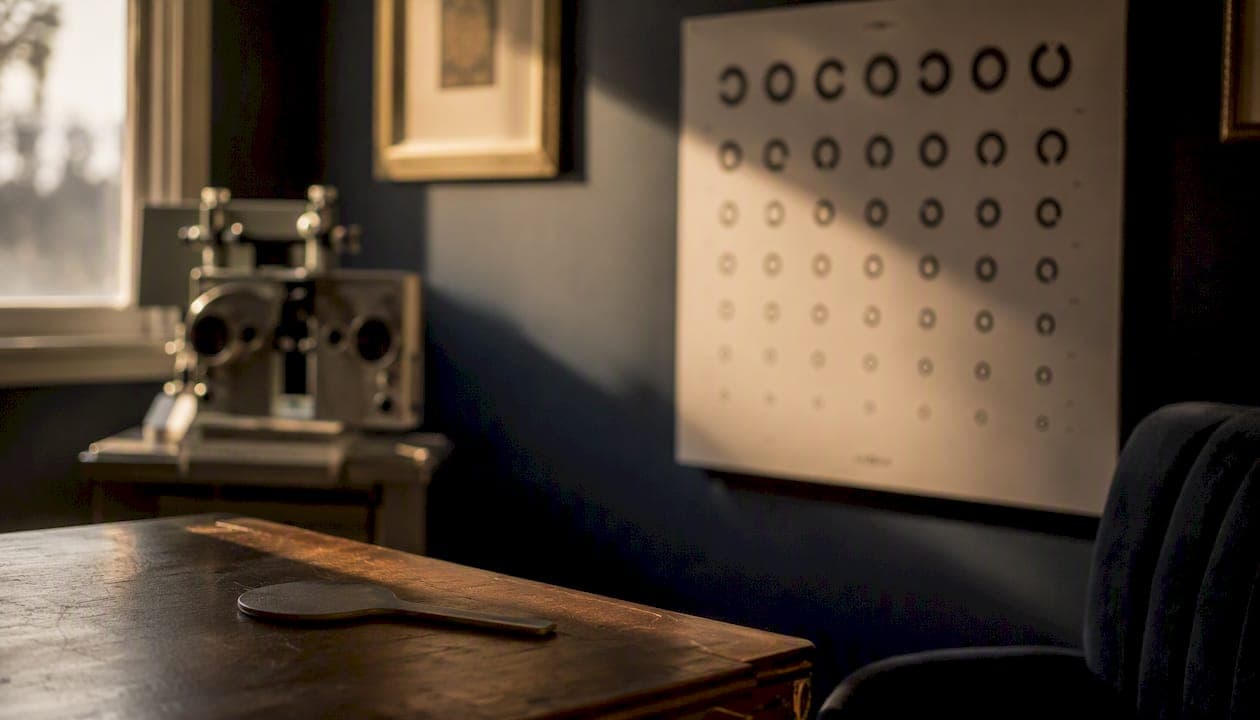
Contrast sensitivity impairment is a reduced ability to detect objects when the difference in brightness or color between an object and its background is small. It's not the same as blurry vision. You can have 20/20 acuity on a Snellen chart and still struggle to see a gray curb against pale pavement at dawn. If everyday tasks like night driving, reading in dim light, or recognizing faces in low-contrast settings have become harder, the right first step is to schedule a comprehensive eye exam and specifically ask your eye care provider about contrast sensitivity testing.
Table of Contents
- What contrast sensitivity impairment actually means for your vision
- Symptoms that suggest your contrast sensitivity is affected
- Common causes and medical conditions linked to reduced contrast sensitivity
- How contrast sensitivity is tested in a clinical setting
- What your contrast sensitivity score actually means
- Treatment options and practical management strategies
- When to see a doctor and what to expect at the appointment
- Research highlights that clinicians and patients should know
- Why contrast sensitivity awareness matters for learning and daily access
- What the evidence on contrast sensitivity impairment tells you to do next
- Primary sources and further reading
- Sources
What contrast sensitivity impairment actually means for your vision
Standard visual acuity testing, the black letters on a white chart, measures how well you resolve fine detail at high contrast. Contrast sensitivity (CS) measures something different: your visual system's ability to detect objects across a range of brightness differences, from stark black-on-white down to barely-there gray-on-gray.
The contrast sensitivity function (CSF) describes how sensitivity varies across spatial frequencies, which is a way of measuring how fine or coarse a pattern is. Sensitivity peaks around 2–5 cycles per degree (mid spatial frequencies) and drops off at both ends of the spectrum. That peak range corresponds roughly to the size of faces, text at reading distance, and objects on a road. Diseases or aging that flatten this curve can devastate daily function while leaving the Snellen result untouched.
The table below shows the key differences between the two measures.
| Feature | Visual acuity (Snellen) | Contrast sensitivity |
|---|---|---|
| What it measures | Finest resolvable detail at high contrast | Ability to detect objects at varying contrast levels |
| Typical stimulus | Black letters on white background | Gratings or letters fading from dark to light |
| When it can be normal | Even with significant CS loss | Even with mild acuity reduction |
| Clinical uses | Routine screening, prescription, legal driving standard | Functional vision, disease monitoring, surgical outcomes |
| Score format | Fraction (20/20) | Log units or letter contrast score (e.g., 2.0) |
A person with early glaucoma or cataracts may pass the acuity test and fail the contrast sensitivity test. That gap is exactly why CS testing adds information the Snellen chart cannot.
Symptoms that suggest your contrast sensitivity is affected
The symptoms of impaired contrast sensitivity are often subtle at first, which is part of why people dismiss them. They tend to show up in specific lighting conditions rather than all the time.
- Night driving difficulty: Oncoming headlights create glare and halos; lane markings on wet pavement disappear; pedestrians in dark clothing seem to materialize at the last second.
- Trouble reading in low light: Text on a cream-colored page, a menu in a dim restaurant, or a phone screen in bright sunlight all become harder to parse.
- Reduced face recognition: Recognizing someone in a shaded hallway or under fluorescent lighting becomes unreliable, even when you know the person well.
- Trips and falls: Curbs, steps, and floor-level obstacles blend into their surroundings. A gray step edge on gray concrete is genuinely invisible to someone with significant CS loss.
- Glare and washed-out vision: Bright environments (snow, beach, a sunny parking lot) cause visual "whiteout" rather than just discomfort.
- Difficulty with low-contrast objects: A faint coin on a beige carpet, a light-gray car against a pale sky, or a white plate on a white tablecloth.
WebMD's clinical overview notes that reduced CS may be present even when a Snellen chart shows normal acuity, which is why these symptoms are often dismissed or attributed to "just getting older." They are worth taking seriously.
Common causes and medical conditions linked to reduced contrast sensitivity
Reduced contrast sensitivity is rarely a condition in isolation. It's usually a signal that something else is happening in the eye or visual pathway.
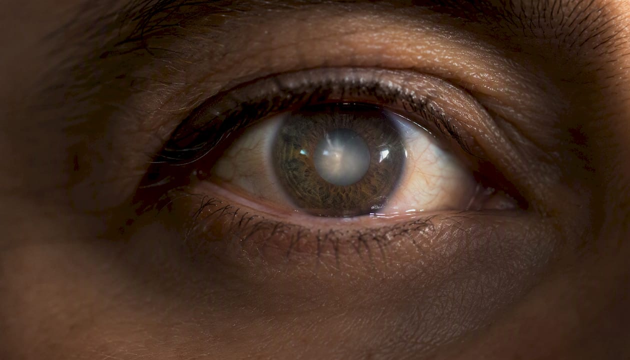
Cataracts are one of the most common causes. The clouded lens scatters incoming light, which reduces contrast before it reaches the retina. CS often declines in cataract patients before acuity does, making it a useful early marker. Cataract surgery typically restores CS substantially.
Glaucoma damages the optic nerve, and CS loss at mid and low spatial frequencies is frequently an early sign, sometimes appearing before measurable visual field loss. Controlling intraocular pressure can slow further decline, but damage already done is generally not reversible.
Age-related macular degeneration (AMD) disrupts the central retina, the region most responsible for fine detail and contrast discrimination. Both dry and wet AMD reduce CS, and the wet form can cause rapid deterioration. Anti-VEGF injections for wet AMD can stabilize or partially improve CS in some patients.
Diabetic retinopathy affects retinal blood vessels and can reduce CS even in its early, non-proliferative stages, before visible hemorrhages appear on imaging. Tight glycemic control is the most effective preventive measure.
Optic neuritis and optic neuropathy inflame or damage the optic nerve directly. Optic neuritis, often associated with multiple sclerosis, frequently causes a dramatic, sudden drop in CS alongside pain with eye movement. Recovery is common but not always complete.
Uncorrected refractive error (nearsightedness, farsightedness, astigmatism) reduces CS by blurring the retinal image. A current, accurate spectacle or contact lens prescription is the simplest fix and is often overlooked.
Beyond diagnosed disease, research in healthy populations shows that age alone increases intraocular light scatter as the lens yellows and thickens, measurably reducing CS even without any pathology. Some systemic medications, including certain antihypertensives and psychotropic drugs, have also been associated with CS changes, though the evidence varies by drug class.
How contrast sensitivity is tested in a clinical setting
CS testing is not automatically included in a routine eye exam. If you have symptoms, ask for it by name. Here is what the main tests involve.
| Test | What it measures | Clinical use | Score format |
|---|---|---|---|
| Pelli–Robson chart | Letter contrast threshold at a fixed size | Broad clinical screening, low-vision assessment | Log contrast sensitivity (0–2.0) |
| Regan/CSV charts | Letter acuity at multiple contrast levels | Functional vision, driving assessment | Log units or contrast percentage |
| Vistech/FACT | Grating contrast across 5 spatial frequencies | Research, disease monitoring, surgical outcomes | Threshold at each frequency band |
| Grating tests | Minimum detectable contrast for sine-wave gratings | Research and specialized clinical use | Contrast threshold (%) |
| Computerized CSF | Full contrast sensitivity function across frequencies | Detailed functional mapping, research | Log sensitivity curve |
The StatPearls point-of-care clinical summary describes all of these tests, their score formats, and their practical interpretation. The Pelli–Robson chart is the most widely used in clinical practice because it is quick (typically 5–10 minutes), requires no special equipment beyond the chart and standard lighting, and produces a single log score that is easy to interpret and track over time.
A few practical points worth knowing before your appointment:
- CS testing is usually done under standardized lighting; bring your current glasses or contacts.
- Each eye is tested separately, and results can differ significantly between eyes.
- Testing is non-invasive and painless, similar in format to a standard acuity test.
- Results can vary slightly between visits due to lighting differences and patient fatigue, so serial testing under consistent conditions matters most for tracking change.
What your contrast sensitivity score actually means
Scores on the Pelli–Robson chart and similar tests are reported in log units. A score of 2.0 is generally considered normal for a young adult. According to StatPearls clinical guidance, a score below 1.5 suggests visual impairment, and a score below 1.0 indicates a visual disability.
Key threshold: Certain Pelli–Robson log contrast sensitivity scores correspond to visual impairment and disability; typical normal scores are known within clinical practice. (StatPearls)
That distinction matters clinically. A patient who scores 1.4 may appear to have only a modest deficit on paper but can struggle significantly with tasks like reading standard print or driving at dusk.
Scores are not perfectly interchangeable between test types. A Pelli–Robson score and a Vistech score describe different things, and clinicians compare results within the same test across visits rather than across test types. If your provider switches tests between appointments, ask them to note which test was used so trends can be tracked accurately.
Treatment options and practical management strategies
Treatment for reduced contrast sensitivity always starts with the underlying cause. There is no medication or supplement that directly restores CS independent of what is causing it.
Medical and surgical treatments:
- Cataract surgery is one of the most effective interventions. Removing the clouded lens and replacing it with a clear intraocular lens typically produces substantial CS improvement, often more dramatic than the acuity improvement alone.
- AMD management: Anti-VEGF injections for wet AMD can stabilize CS and sometimes improve it. Dry AMD has no proven treatment that reverses CS loss, though AREDS2 supplementation (vitamins C and E, lutein, zeaxanthin, zinc) is recommended to slow progression.
- Glaucoma control: Lowering intraocular pressure through drops, laser, or surgery slows further optic nerve damage and CS decline, but does not restore what is already lost.
- Diabetic retinopathy care: Glycemic control, blood pressure management, and timely laser or anti-VEGF treatment reduce the risk of CS deterioration.
Optical and environmental strategies:
Anti-reflective coatings on lenses reduce glare and can improve functional contrast in bright conditions. Yellow or amber tinted lenses increase perceived contrast in some lighting conditions, particularly overcast daylight. However, the American Academy of Ophthalmology cautions against tinted lenses for night driving: they reduce overall light transmission and can distort color perception in ways that create new hazards rather than solving old ones.
Magnification, whether through reading glasses, handheld magnifiers, or screen zoom settings, helps with near tasks by enlarging the image and effectively increasing the contrast signal reaching the retina.
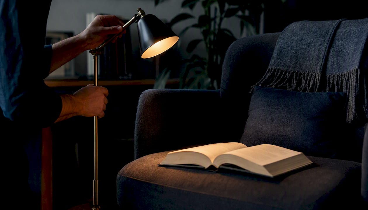
Pro Tip: The most overlooked home adjustment is lighting direction, not just brightness. A lamp positioned to the side of a reading surface (rather than overhead) reduces surface glare and increases the shadow contrast that defines edges and text. Matte surfaces on counters and floors also help: glossy finishes reflect ambient light and wash out the contrast cues that define edges and depth. High-contrast labeling on appliance controls, stair edges, and light switches costs almost nothing and can meaningfully reduce fall risk.
Low-cost home modifications, including higher directional lighting, high-contrast labels, and matte surfaces, can improve day-to-day function while clinical evaluation and treatment proceed.
When to see a doctor and what to expect at the appointment
Some symptoms warrant prompt evaluation; others can be addressed at a scheduled routine visit. Here is how to tell the difference.
Seek urgent evaluation (within days) if you notice:
- Sudden loss of contrast or vision in one or both eyes
- New visual distortion (straight lines appearing wavy or bent)
- Severe, sudden glare or light sensitivity that is new
- Difficulty driving that developed rapidly rather than gradually
Schedule a routine comprehensive exam if you have:
- Gradual difficulty with night driving or dim-light reading
- Increasing trouble recognizing faces in low-contrast settings
- A history of cataracts, glaucoma, AMD, or diabetes with new visual complaints
- Symptoms that your standard acuity test has not explained
At a contrast-sensitivity-focused exam, expect the following sequence. Your provider will take a detailed history about when symptoms started, which tasks are affected, and whether they are worse in specific lighting. A standard high-contrast acuity test comes first, followed by contrast sensitivity testing (most likely Pelli–Robson). A slit-lamp exam checks the anterior segment, including the lens for cataract. Retinal and optic nerve evaluation follows, often with dilation. Depending on findings, your provider may order optical coherence tomography (OCT) imaging, visual field testing, or refer you to a retinal specialist or neuroophthalmologist.
An optometrist can perform the full CS workup and manage many underlying conditions. An ophthalmologist (or subspecialist) is appropriate when surgical intervention, retinal disease, or neurologic involvement is suspected. If your optometrist finds something concerning, they will refer you directly.
Pro Tip: CS testing is not always included in a routine eye exam. StatPearls clinical guidance recommends that symptomatic patients specifically request it. Before your appointment, write down the specific situations where your vision feels inadequate, because a detailed symptom history is what prompts the right tests.
Research highlights that clinicians and patients should know
A few evidence-backed findings are worth having in front of you when you talk to your provider.
- CS often declines before acuity in cataract patients. This makes CS a more sensitive early marker of lens-related visual decline than the Snellen chart alone. (StatPearls)
- The CSF peaks around 2–5 cycles per degree. This mid-frequency range corresponds to the visual tasks most people care about: reading, face recognition, and road navigation. Diseases that flatten the CSF at this range have outsized real-world impact. (NCBI contrast sensitivity function chapter)
- CS adds information that acuity misses. Multiple studies confirm that CS deficits can be present with normal Snellen results, and that CS scores correlate more strongly with functional complaints (driving difficulty, reading problems) than acuity scores do. (WebMD clinical overview)
- Age and lens changes reduce CS in healthy people. Increased intraocular light scatter from a yellowing lens measurably reduces CS even without diagnosed disease, which is why CS tends to decline gradually from middle age onward. (PMC factors study)
- CS loss at specific spatial frequencies points to specific diseases. Glaucoma tends to affect mid and low frequencies; optic neuritis often affects high frequencies first. Knowing which band is affected helps clinicians narrow the differential diagnosis. (NCBI contrast sensitivity function chapter)
Research note: A CS score of 2.0 is typically normal; below 1.5 suggests impairment; below 1.0 indicates disability. Because the scale is logarithmic, each 0.5-unit drop represents a tenfold change in sensitivity. (StatPearls)
Why contrast sensitivity awareness matters for learning and daily access
Contrast sensitivity impairment affects far more than driving. For many people, it quietly erodes the ability to read, learn, and engage with written content, often years before a diagnosis is made. If standard print feels harder to process, if screens wash out in bright light, or if low-contrast digital text causes fatigue, the problem may be partly visual rather than cognitive.
Coreforgeaudio exists precisely for moments like this. The platform is built for people whose access to written content is limited by visual impairments, reading differences, or other barriers. Human-narrated audiobooks remove the contrast requirement entirely: there is no page, no font, no lighting condition to manage. For anyone navigating a CS diagnosis or waiting for treatment to take effect, audio-based reading access can keep learning and engagement going in the meantime.
If you are supporting someone with a visual impairment, the Coreforgeaudio blog also covers how visually impaired users access digital content and audio description platforms for visually impaired readers, both of which offer practical strategies beyond what a single eye exam can address.
Schedule a comprehensive eye exam. Ask specifically about contrast sensitivity testing. And while you wait, consider whether an audio-first reading format could reduce the daily strain.
What the evidence on contrast sensitivity impairment tells you to do next
Contrast sensitivity impairment is a measurable, clinically significant reduction in the ability to detect objects at low contrast, and it is frequently missed by standard acuity testing alone.
| Point | Details |
|---|---|
| CS differs from acuity | A 20/20 Snellen result does not rule out meaningful contrast sensitivity loss. |
| Key clinical thresholds | Scores below 1.5 suggest impairment; scores below 1.0 indicate visual disability. |
| Common causes | Cataracts, glaucoma, AMD, diabetic retinopathy, and optic neuritis are the most frequent culprits. |
| Request the test | CS testing is not always included in routine exams; ask for it by name if symptoms are present. |
| Practical home changes | Directional lighting, matte surfaces, and high-contrast labels reduce fall risk while treatment proceeds. |
Primary sources and further reading
The following sources were used in this article. The first three are peer-reviewed clinical references; the fourth is a patient-facing clinical summary; the fifth is a research study in healthy populations.
- Contrast Sensitivity — StatPearls, NCBI Bookshelf — Peer-reviewed clinical reference covering definition, causes, tests, and numeric thresholds.
- Contrast Sensitivity Function — NCBI Bookshelf — Clinical chapter on the CSF, spatial frequency concepts, and measurement scales.
- Contrast Sensitivity: Treatment & Management — StatPearls Point of Care — Clinical point-of-care summary with test descriptions, score formats, and interpretation guidance.
- Contrast Sensitivity Issues With Vision — WebMD — Patient-facing overview of symptoms, real-world impacts, and when to seek care.
- Factors Affecting Contrast Sensitivity in Healthy Individuals — PMC — Peer-reviewed research study on age, lens changes, and CS decline in people without diagnosed eye disease.
This article is general information, not a substitute for advice from a qualified doctor. Consult a qualified healthcare professional about your own circumstances before acting on anything here.
Sources
- Contrast Sensitivity - StatPearls - NCBI Bookshelf
- CONTRAST SENSITIVITY FUNCTION
- What Are Contrast Sensitivity Issues With Vision? - WebMD
- Factors Affecting Contrast Sensitivity in Healthy Individuals
- Contrast Sensitivity | Treatment & Management | Point of Care - StatPearls